The AKT1 Polyclonal Antibody (CAB11016) is a valuable tool for researchers studying AKT1, a key protein involved in various cellular processes such as cell survival, proliferation, and metabolism. This antibody, generated in rabbits, exhibits high specificity and sensitivity towards human samples, making it ideal for use in Western blot analyses.AKT1, also known as protein kinase B, plays a crucial role in regulating signaling pathways that control cell growth and survival. Dysregulation of AKT1 has been implicated in various diseases, including cancer, diabetes, and neurological disorders.
By utilizing the AKT1 Polyclonal Antibody, researchers can accurately detect and analyze AKT1 expression in different cell types, providing valuable insights into its function and potential as a therapeutic target.Overall, the AKT1 Polyclonal Antibody is a reliable tool for investigating the role of AKT1 in cellular processes and disease pathogenesis, offering researchers a valuable resource in advancing their studies in cancer research, metabolic disorders, and other related fields.
Product Name:
AKT1 Polyclonal Antibody
SKU:
CAB11016
Size:
20uL, 100uL
Isotype:
IgG
Host Species:
Rabbit
Reactivity:
Human,Mouse,Rat
Immunogen:
A synthetic peptide corresponding to a sequence within amino acids 350-480 of human AKT1 (NP_005154.2).
This gene encodes one of the three members of the human AKT serine-threonine protein kinase family which are often referred to as protein kinase B alpha, beta, and gamma. These highly similar AKT proteins all have an N-terminal pleckstrin homology domain, a serine/threonine-specific kinase domain and a C-terminal regulatory domain. These proteins are phosphorylated by phosphoinositide 3-kinase (PI3K). AKT/PI3K forms a key component of many signalling pathways that involve the binding of membrane-bound ligands such as receptor tyrosine kinases, G-protein coupled receptors, and integrin-linked kinase. These AKT proteins therefore regulate a wide variety of cellular functions including cell proliferation, survival, metabolism, and angiogenesis in both normal and malignant cells. AKT proteins are recruited to the cell membrane by phosphatidylinositol 3,4,5-trisphosphate (PIP3) after phosphorylation of phosphatidylinositol 4,5-bisphosphate (PIP2) by PI3K. Subsequent phosphorylation of both threonine residue 308 and serine residue 473 is required for full activation of the AKT1 protein encoded by this gene. Phosphorylation of additional residues also occurs, for example, in response to insulin growth factor-1 and epidermal growth factor. Protein phosphatases act as negative regulators of AKT proteins by dephosphorylating AKT or PIP3. The PI3K/AKT signalling pathway is crucial for tumor cell survival. Survival factors can suppress apoptosis in a transcription-independent manner by activating AKT1 which then phosphorylates and inactivates components of the apoptotic machinery. AKT proteins also participate in the mammalian target of rapamycin (mTOR) signalling pathway which controls the assembly of the eukaryotic translation initiation factor 4F (eIF4E) complex and this pathway, in addition to responding to extracellular signals from growth factors and cytokines, is disregulated in many cancers. Mutations in this gene are associated with multiple types of cancer and excessive tissue growth including Proteus syndrome and Cowden syndrome 6, and breast, colorectal, and ovarian cancers. Multiple alternatively spliced transcript variants have been found for this gene.
Purification Method:
Affinity purification
Gene ID:
207
Storage Buffer:
Store at -20℃. Avoid freeze / thaw cycles.Buffer: PBS with 0.05% proclin300,50% glycerol,pH7.3.
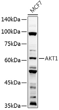
Western blot analysis of lysates from MCF7 cells, using AKT1 Rabbit pAb (CAB11016) at 1:1000 dilution.Secondary antibody: HRP Goat Anti-Rabbit IgG (H+L) (CABS014) at 1:10000 dilution.Lysates/proteins: 25μg per lane.Blocking buffer: 3% nonfat dry milk in TBST.Detection: ECL Basic Kit (AbGn00020).Exposure time: 60s.

")
")
")
")
")
")
")